HOME
Medical Topics:
Ligament
Injury
Disease
About The Doctor:
Questions?
Email at:
GNYaco@aol.com
| ARTICULAR
CARTILAGE INJURY |
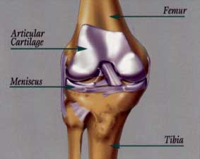 |
1. Definition: Articular or Hyaline cartilage covers the end of
the long bones and forms the joint surfaces. It is a unique
structure in that it can withstand enormous compressive
forces and create a low friction surface for the joint to
glide on. This complex organ is made up of cells called
chondrocytes and matrix composed of proteins and sugars
in a specific interwoven fabric.
2. Injury: Articular cartilage is injured when
the knee joint is
compressed
under heavy load or when angular or shear
forces
are applied to the surface. The result is one of
several
possible lesions...softening, fissuring, fragmenting
or complete
removal of the cartilage covering. Symptoms
include
pain, swelling and subsequent loss of joint function.
Adult articular
cartilage does not repair itself. The reason
is that
the chondrocytes have little mobility and there is no
blood supply
to the matrix to provide healing elements. The
above mentioned
lesions then, are more or less permanent.
Worse yet,
they progress from softening to complete
destruction
of the joint if left untreated.
TREATMENT: If there are correctable causes of
the articular
cartilage
problem, these must be addressed first:
a) Loss of meniscus - early stages of cartilage damage
due to
loss of meniscal tissue and subsequent high
compressive
loads are addressed by meniscal allograft
reconstruction
(see Meniscus).
|
b) Malalignment - abnormal knee
alignment is corrected by distal femoral or proximal tibial osteotomy. This is where a wedge of bone is surgically added to or removed from the bone adjacent to the knee to recreate a horizontal joint line and eliminate high angular stresses on the cartilage. |
 |
(ACL) creates gliding movements in the joint and high
shear stress on the articular cartilage. This is treated with
ACL reconstruction (see Anterior Cruciate Ligament).
Once these causes are corrected, the articular cartilage
lesions can be addressed. Symptomatic areas of fragmented
or destroyed cartilage have traditionally been treated by
abrading or puncturing the underlying bone. These
techniques lead to healing with fibrocartilage which does
not possess the same mechanical properties as hyaline
cartilage and lasts only 10 to 12 months. Newer technologies
are aimed at repairing these defects with tissue that
reproduces the original hyaline cartilage surface. These
innovative treatments include:
----
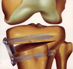
b) Osteochondral Autograph Transfer (OATS) - smaller
Dr.Yacobucci
has performed over 20 of these procedures
Dr. Yacobucci is one of only a few knee surgeons in

Click to see enlarged picture
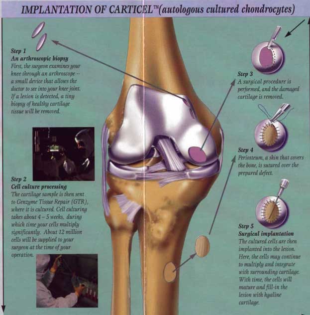
a) Autologous Chondrocyte Implantation (carticel) - if a
full
thickness defect in the articular cartilage (>2 cm.
diameter)
is found in a weight bearing area of the femoral
condyle,
a biopsy is obtained arthroscopically from a
non-functional
area of the same knee. This sample is sent to
a laboratory
where the cartilage cells are manipulated to
replicate
and produce hyaline like matrix. These
"stimulated"
cells are suspended in a liquid medium and
then injected
into the prepared defect during the second
(open)
surgery. The defect will subsequently fill with this
new cartilage
and the patients symptoms will improve as
the cartilage
matures over 1 year. The cost of this complex
laboratory
process is over $11,000.00...however this is more
then justified
if it saves a young, healthy, productive person
from prolonged
disabling knee pain, slow deterioration of
the joint
and ultimately a knee replacement at a relatively
young age.
This procedure is only recommended for
patients
who are between the ages of 15 and 55 and have an
otherwise
normal knee (alignment, meniscus, and ligament).
Dr. Yacobucci
has flown to Sweden to receive special
training
in this technique from the surgeon who pioneered
this operation.
He has performed over 20 of these
surgeries
and is actively involoved in clinical research and
teaching as it
pertains to this complex procedure.
full
thickness defects can be treated by transferring punch
grafts
of bone and cartilage from a healthy non-functional
area in
the knee into the prepared defect. The advantages
over carticel
are this can be done during one arthroscopic
procedure,
and the cost is significantly less. This technique
is quite
demanding as positioning of the grafts must be
precise
to create a smooth, functional articular
cartilage
surface.
There are no age limits on patient selection and the
condition
of the remainder of the joint need not
be perfect.
Full maturation of the graft is complete at about
12 weeks
post-op.
and has
given numerous seminars about this innovative
operation.
![]()
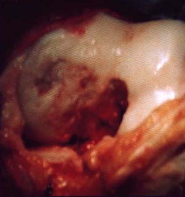
![]()
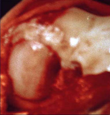
Before
After
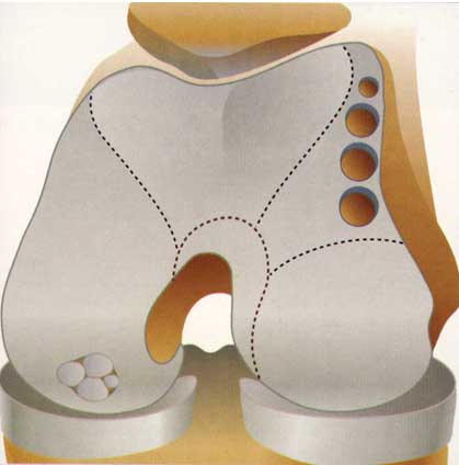
c) Osteoarticular Allograft Reconstrucion - a new
technique
of harvesting, testing and preserving bone and
articular
cartilage from deceased human tissue donors
allows
us to use these large (up to 4 cm. diameter) grafts to
fill massive
femoral condyle surface defects. This is the
result
of a patented technique developed by Cryolife
corporation
that preserves large numbers of viable
chondrocytes
in the grafted tissue. Advanced
instrumentation
is used to exactly duplicate the contour of
the cartilage
surface being repaired. A press fit secures the
graft at
the recipient site. The cost of tissue and
intrumentation
is $10,000 however, this procedure has the
potential
of saving a knee joint from gradual complete
destruction.
Arizona
who have successfully carried out this procedure.
Back to the Top